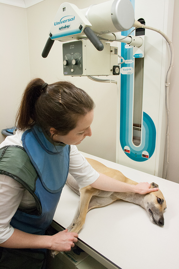
Our Surgery is fully equipped to take X-Rays (often called Radiographs) of your pets. Radiographs are a very important tool to help us diagnose diseases in animals particularly for conditions involving bones, chest or abdomen. Our veterinarians will discuss your pet’s case and conduct a thorough health check to determine if your pet requires radiography. In many cases where you think it may be necessary to have radiographs, our experienced vets can diagnose and treat conditions due to the years of working with pets.
Most of our patients are admitted to the hospital for the day to have radiographs taken. In an emergency, we will take them immediately. We ask that you bring your pet in unfed on the morning of admission, as they will most likely be sedated or anaesthetized to allow us to take the best quality radiographs possible. The images are developed within minutes and we can read the results straight away.
Why do pets need to be sedated or anaesthetized to have Radiographs taken?
When we have radiographs taken the radiographer asks us to keep perfectly still, often in unnatural positions. Most pets would never lie still enough for us to take the quality radiographs required to diagnose their condition. Sedation and anaesthesia allow us to get the most useful radiographs possible.
How are X-Rays made?
Taking a radiograph is very similar to taking a photo, except we use X-Rays instead of light rays. The usefulness of radiography as a diagnostic tool is based upon the ability of X-Rays to penetrate matter, Different tissues in the body absorb X-Rays to differing degrees.Of all the tissues in the body, bone absorbs X-Rays the best. This is the reason bone appears white on a radiograph whereas air appears black because it absorbs no X-Rays. Soft tissues such as intestine and organs absorb some but not all X-Rays. We will demonstrate and explain the results of your pet’s radiographs when they go home.